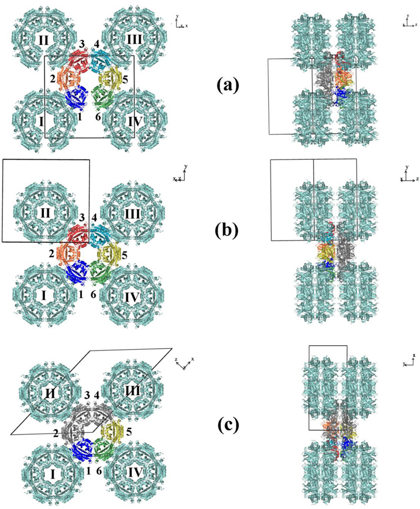
Fig. 2.
Crystal packing. Molecular layers in the structure of SpeG in open dodecameric state (a), intermediate dodecameric state (b) and closed dodecameric state (c). The monomers of the asymmetric unit cell are colored. The 2-fold rotation axis goes along the crystallographic axis Y. Monomers of the SpeG dodecamer that are related by crystallographic symmetry are shown in grey. The GNAT dimers in the dodecameric structure that are composed from monomers related by 2-fold crystallographic or noncrystallographic rotation axis are numbered clockwise from 1 to 6.

