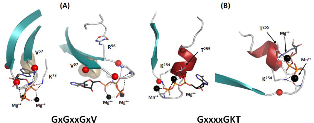
Figure 2.
G-loops and P-loops. The G-loop of PKA (Protein Data Bank[105] identifier (PDBID): 1ATP) is compared to the P-loop of Phosphoenolpyruvate Carboxykinase (PCK) (PDBID: 1AQ2). (A) The G-loop connects two β-strands and contains three glycines (red spheres). The residue that follows the last glycine points away from the ATP phosphates. Right after this residue there is a highly conserved valine that is a part of the C-spine (tan surface). (B) P-loop connects a β-strand and an α-helix and contains two conserved glycines (red spheres) that are followed by lysine and serine/threonine. Unlike Arg56 and Val57 in PKA Thr255 and Lys254 in PCK contribute directly to metal and phosphate binding.

