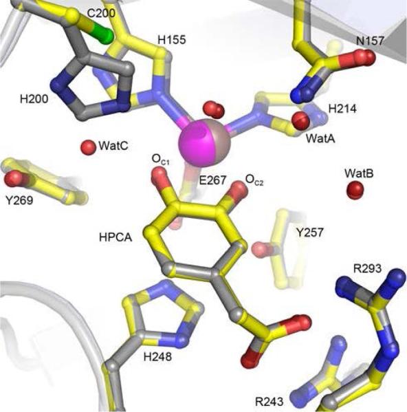
Figure 2.
Comparison of active sites of the WT HPCD and the H200C variant in complex with HPCA. Structure overlay of the WT HPCD (PDB 4GHG) and H200C (PDB 5BWH) structures. Atom color code: gray, carbon (HPCD); yellow, carbon (H200C); dark blue, nitrogen (HPCD); blue, nitrogen (H200C); dark red, oxygen (HPCD); red, oxygen (H200C); green, sulfur (H200C); bronze, iron (HPCD); purple, iron (H200C). Cartoons depict secondary structure elements for the H200C variant (gray) and HPCD (light blue). WatA-C represent crystallographically observed (not metal-coordinated) solvent in the active site.

