
Figure 2.
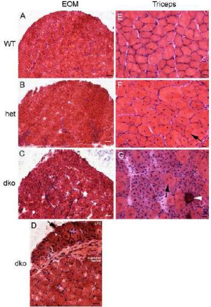
Sparing of the Extraocular Muscles (EOM) in mdx:utrophin+/− (het) and mdx:utrophin−/− (dko) Mice but not the Levator Palpebrae Superioris Muscle or Triceps Skeletal Muscles. Photomicrographs of muscle specimens stained with hematoxylin and eosin obtained from mice at 3 months of age, except the mdx:utrophin−/− muscles which were obtained from 2 month old mice. Extraocular muscles from A. a wild type (WT) control EOM, B. an mdx:utrophin+/− (het) EOM, and C. an mdx:utrophin−/− (dko) EOM demonstrate that there was little sign of muscle pathology in the mouse models of muscular dystrophy. Bar for A-C is 100 μm. D. Photomicrograph of the levator palpebrae superioris (LPS) and the superior rectus muscle of an mdx:utrophin−/− (dko) mouse. Note that the LPS was significantly affected, with central nucleation (arrow) in almost every myofiber present, while the superior rectus was normal in appearance. E. Photomicrographs of normal triceps muscle from a wild type (WT) mouse, F. an mdx:utrophin+/− (het) triceps muscle, and G. an mdx:utrophin−/− (dko) triceps muscle demonstrate dystrophic pathology. There were many centrally nucleated myofibers in the mdx:utrophin+/− limb muscle specimen. The pathology was even more in the triceps muscle from the dko mouse. This included basophilic myofibers suggestive of new regeneration and accelerated protein synthesis, central nucleation (arrow), and a calcified fiber (arrowhead). For D-G, bar is 20μm.