Figure 6.

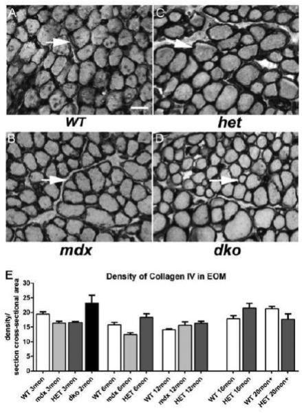
Density of Collagen IV in WT, mdx, mdx:utrophin+/− (het), and mdx:utrophin−/− (dko) Mice. Photomicrographs of collagen IV immunostaining in the EOM of WT (A), mdx (B), mdx:utrophin+/− (het) (C), and mdx:utrophin−/− (dko) mice (D). Arrows indicate collagen IV around individual myofibers. The collagen IV levels in the disease models did not appear to differ significantly from levels in WT control mice. Bar is 50μm. E. Quantification of collagen IV density in the EOM of WT, mdx, mdx:utrophin+/−, and mdx:utrophin−/− at indicated intervals show that there were no significant changes in the level of collagen IV in the four genotypes examined. Data are expressed as mean ± SEM.
