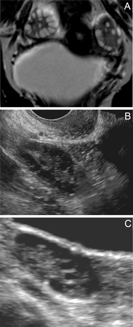
Figure 1.
Ovarian morphology. (A) MRI, (B) Transvaginal US, (C) Transabdominal US. Figure 1A displays a coronal view by MRI of an ovary in an adolescent subject with PCOS. Follicles (hyperintense) are clearly demarcated from stroma (hypointense). Figures 1B and 1C are ultrasound images from adolescent subjects with PCOS, with Figure 1B representing a transvaginal image and 1C representing a transabdominal image. Follicles are visualized in black (hypoechoic) with stroma appearing more hyperechoic. Distinguishing individual follicles by ultrasound is difficult, precluding a follicle count.

