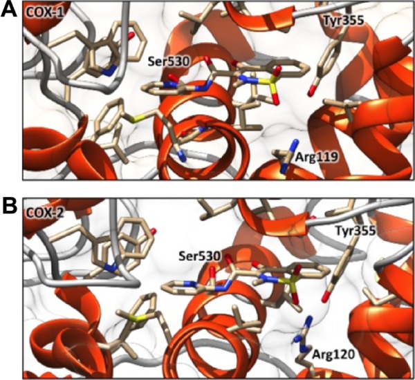
Figure 2.

Best docking complexes between COX-1 (A), COX-2 (B), and PXM.
Notes: The α-helices are shown as orange spirals and the loops are represented by light gray wires. The PXM molecules, hosted in both the active sites in a very similar binding mode, are depicted by stick representations.
Abbrevaitions: COX-1 and -2, cyclo-oxygenases-1 and 2; PXM, piroxicam.
