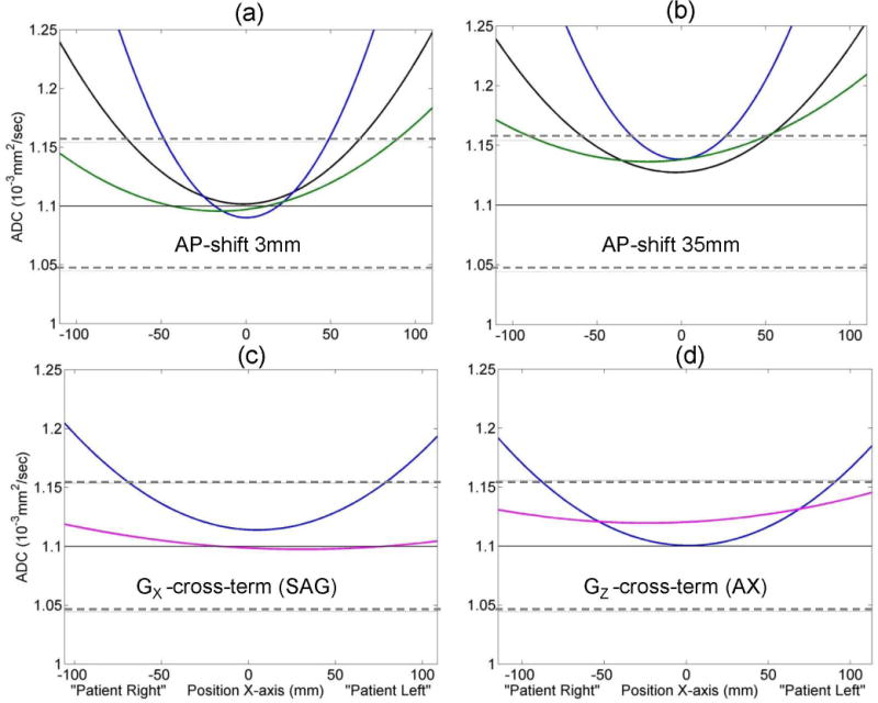
Figure 2.

Vertical offset in measured ADC value is illustrated for individual system gradient channels (GX: blue, GY: green) and the trace (black) due to phantom positioning error (AP-elevation) in (a) versus (b), and due to cross-terms between diffusion and imaging gradients on slice-select channel (GX: blue, GZ: magenta) for sagittal (c) versus axial (d) scans. Solid horizontal lines mark the unbiased (known) ice-water ADC value. Dashed horizontal lines mark ±5% deviation. Head first, supine positioning was used for “Patient RL” direction assignment.
