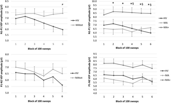
Fig. 2.

Raw amplitudes (mean ± SEM) of N1-P1 (upper graphs) and P1-N2 (lower graphs) VEP components in 6 sequential blocks of 100 recordings. On the left healthy volunteers [HV, n = 30] are compared to the total group of migraine with aura patients [MAtot, n = 47]; on the right they are compared to the 2 subgroups of patients with pure visual aura [MA, n = 27] and patients with complex aura [MA+, n = 20]. ≠ p < 0.05 MAtot vs HV; *p < 0.05 MA+ vs MA; § p < 0.05 MA+ vs HV
