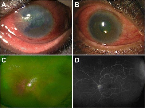
Fig. 1.

A case of bilateral tubercular endogenous endophthalmitis with scleritis. a Slit lamp biomicroscopy of the left eye with diffuse and circumcorneal congestion and scleral involvement. There is corneal edema and opacification superiorly. The pupil has broad-based synechiae, and the view of the posterior segment was hazy. b The right eye with severe congestion and ciliary injection. There was a yellow glow present (visible near the inferior pupillary border). c A wide-angled fundus photograph of the left eye with vitreous haze secondary to vitritis along with focal sheathing of superior vessels. The fluorescein angiography (d) shows presence of superior perivascular hyperfluorescence and leakage of dye in the superotemporal periphery
