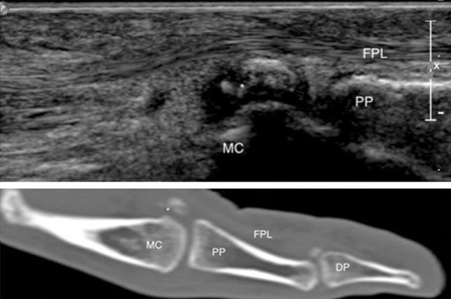
Fig. 4.

Longitudinal US scan (on top) with corresponding CT image (below) over the ulnar sesamoid of the MCP joint of the thumb demonstrates the fragments (asterisk) of the sesamoid. MC first metacarpal, PP proximal phalanx, DP distal phalanx, FPL flexor pollicis longus
