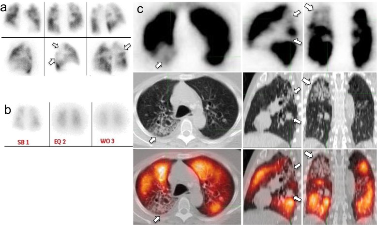
Fig. 1.
Nondiagnostic V/Q scan interpreted as negative for PE on Q-SPECT/CT. Mismatched perfusion defects (arrows, panel a: planar Q scan; panel b: posterior planar V scan with Xenon, which was performed after the planar Q scan) would be read as intermediate probability for PE according to the PIOPED II criteria, but correlate with branching ground-glass opacities on Q-SPECT/CT images (arrows, panel b, top row: Q-SPECT; mid row: noncontrast LDCT; lower row: fusion Q-SPECT/CT), consistent with inflammatory/infectious change, thus interpreted as negative for PE

