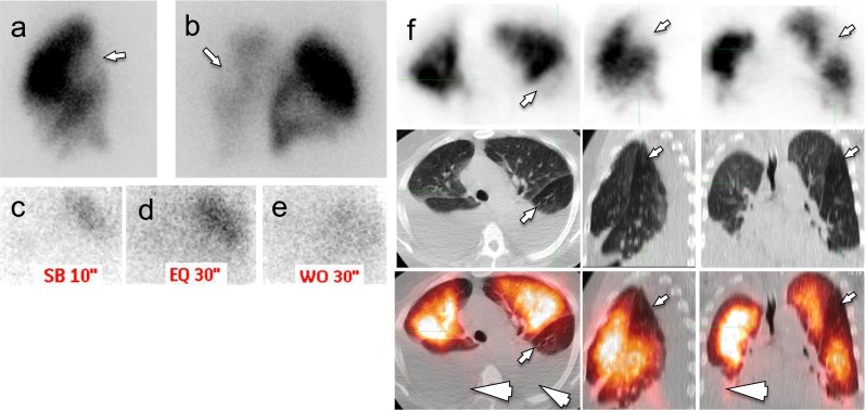
Fig. 5.

False-positive Q SPECT/CT. Nondiagnostic planar V/Q scan showed perfusion defects in the left lung (arrows, a right lateral planar Q scan; b right posterior oblique planar Q scan; limited planar V scan with Xenon on right posterior oblique view after planar Q scan; c inspiration phase; d equilibrium phase; e wash-out phase). On Q-SPECT/CT images (panel f, top row: Q-SPECT; mid row: noncontrast LDCT; lower row: fusion Q-PECT/CT), the perfusion defects (small arrows) appeared without a corresponding CT abnormality and was read as positive for PE. Additional bilateral pleural perfusions were also noted (arrowheads). A CTPA obtained 3 days later showed a patent pulmonary artery branch in this region. The case is deemed false positive for Q-SPECT/CT
