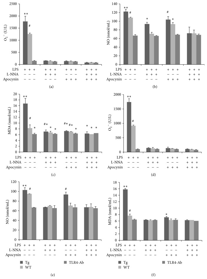
Figure 4.
Overexpression of TLR4 induced oxidative stress via the secretion of NO by monocytes/macrophages. Levels of O2 −, NO, and MDA were examined in monocytes/macrophages after LPS stimulation. Cells were treated with various combinations of the inhibitors apocynin (10 mmol/L) and L-NNA (10 mmol/L; (a), (b), and (c)). Similar patterns were observed when inhibitor concentrations were raised to 20 mmol/L ((d), (e), and (f)). Tg: transgenic sheep, WT: wild-type, and TLR4-Ab: anti-TLR4 antibody. The results are expressed as mean ± SE. ∗, ∗∗, #Values were found to be significantly different between groups (P < 0.05).

