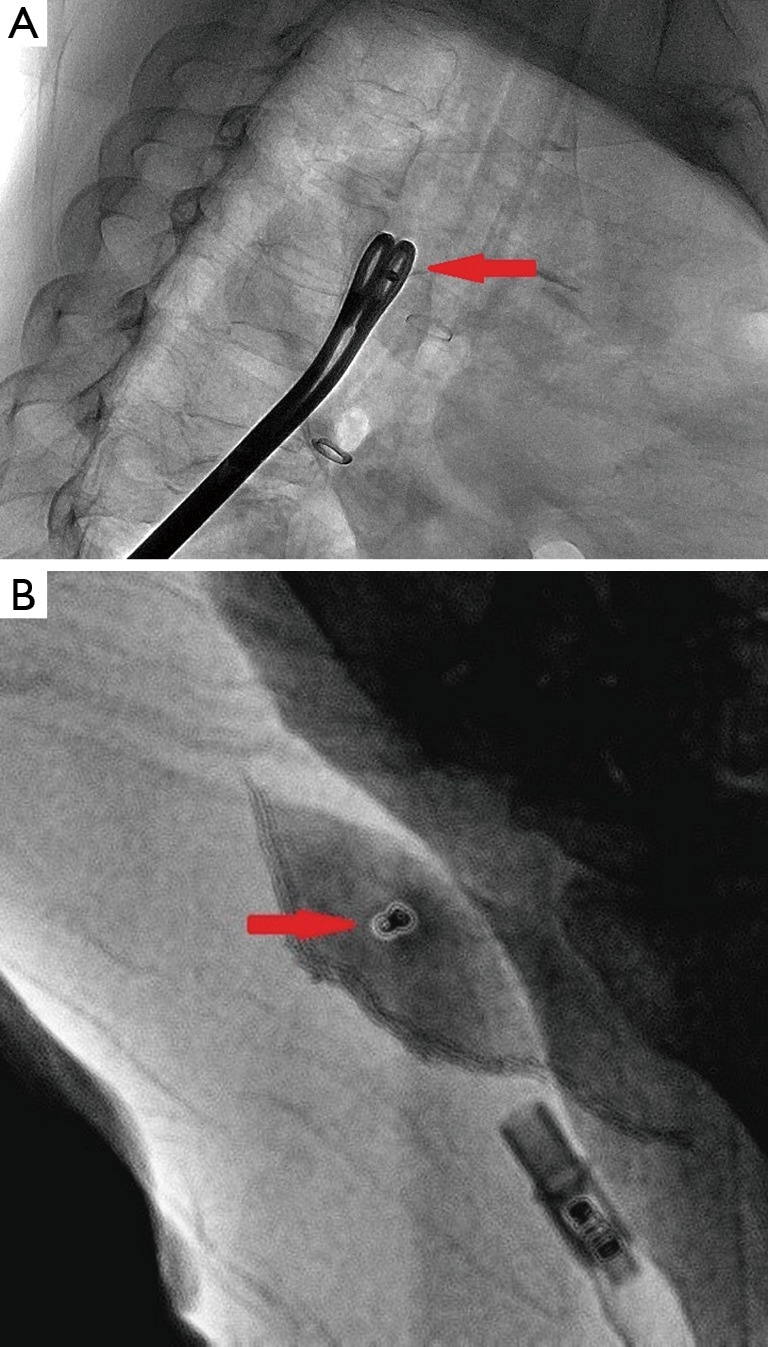
Figure 3.

(A) Real-time digital subtraction angiography (DSA) is used to ensure the placement of the coil (red arrow) and to accurately grasp the surrounding lung tissues; (B) the coil is visualized in the specimen, and the range is >2 cm from the incisional margin to the solitary pulmonary nodules (SPNs).
