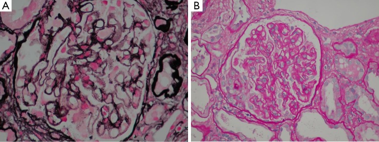
Figure 1.

Kidney biopsy specimen. (A) Silver staining with an optical microscope reported spiculated projections at a right angle with respect to the basement membrane “spikes”. The immune deposits were not argyrophilic and were observed as clear spaces between the “Spikes”. (B) Hematoxylin-eosin stain using an optical microscope, showing a necrotizing crescentic glomerulonephritis induced by inflammation and diffuse thickening of basement membranes with stiffness (evident at the top of the sphere). Magnification: A, 400×; B, 200×.
