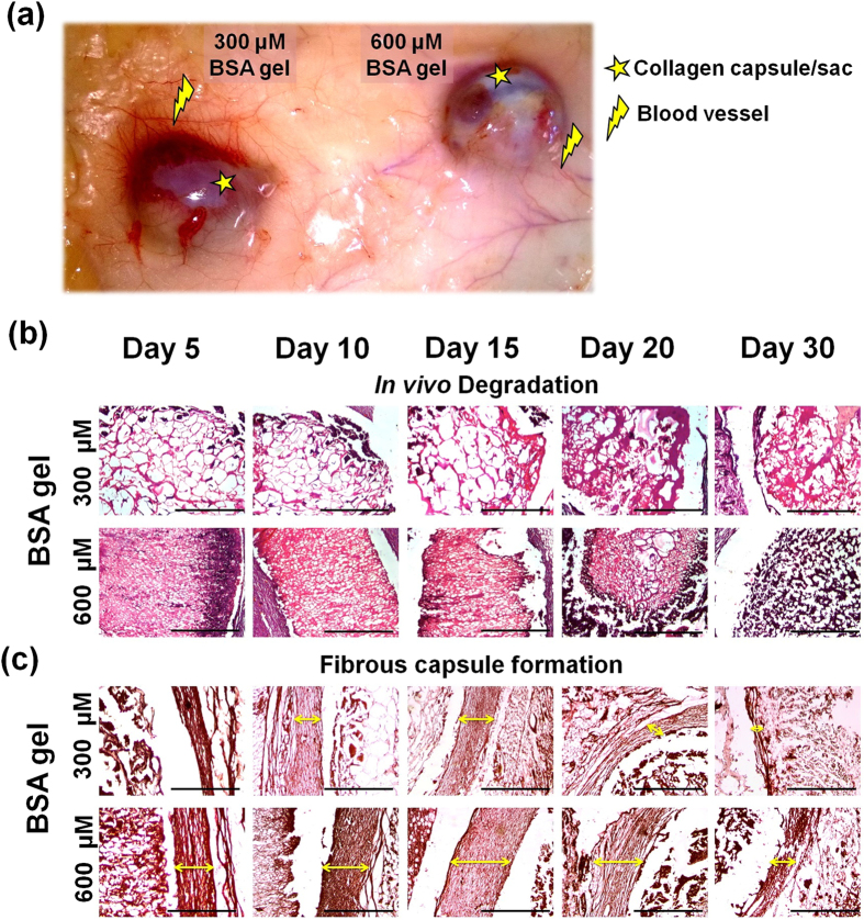
Figure 3. In vivo degradation and tissue response analysis of BSA gel indented as a subcutaneous implant in albino rats.
(a) Explants of BSA gel obtained on day 20, showing lesser collagen capsule and increased blood vessels for 300 μM BSA gel compared to 600 μM BSA gel (b) In vivo biodegradation assessment of BSA gel prepared at 300 μM (bigger pore size) and 600 μM (smaller pore size) concentration. (c) Observations on the fibrous capsule formation around the BSA gel.

