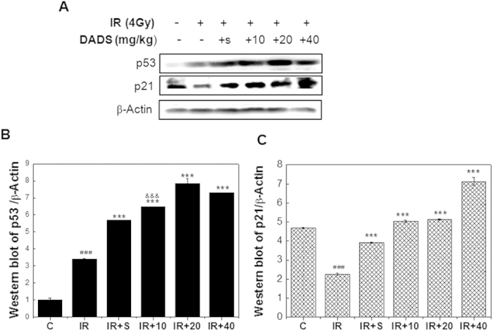
Figure 3. Effect of DADS on the protein levels of p53 and p21 in carbon ion–irradiated mouse testis.

(A) Representative western blot images. (B,C) Quantitative analysis of p53 and p21 protein in mouse testis tissues by western blot analysis. β-Actine was used as a loading control. Relative expression of different protein compaired with control in the same time. Values represent the average ± SD from three gels per group. ###P < 0.001 versus the control group for IR group; ***P < 0.001 versus the IR group for the group received DADS for 3 days prior to carbon ion irradiation.
