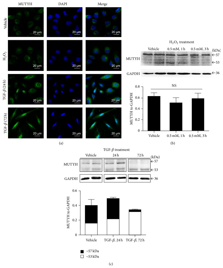
Figure 2.
In vitro regulation of MUTYH in HK-2 tubular cells treated with H2O2 and TGF-β1. Immunofluorescent images demonstrated the location of MUTYH (in green) compared to DAPI (in blue) staining (a). Immunoblot analysis showed similar expression of MUTYH after H2O2 treatment (b) and distinct regulation of MUTYH after TGF-β1 treatment (c).

