Figure 2.

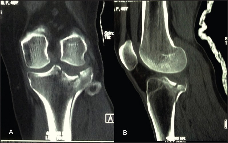
CT scan, (a) coronal view showing the depression of the articular surface and (b) sagittal view demonstrating the posterior placement of the fracture site

CT scan, (a) coronal view showing the depression of the articular surface and (b) sagittal view demonstrating the posterior placement of the fracture site