
Figure 3.
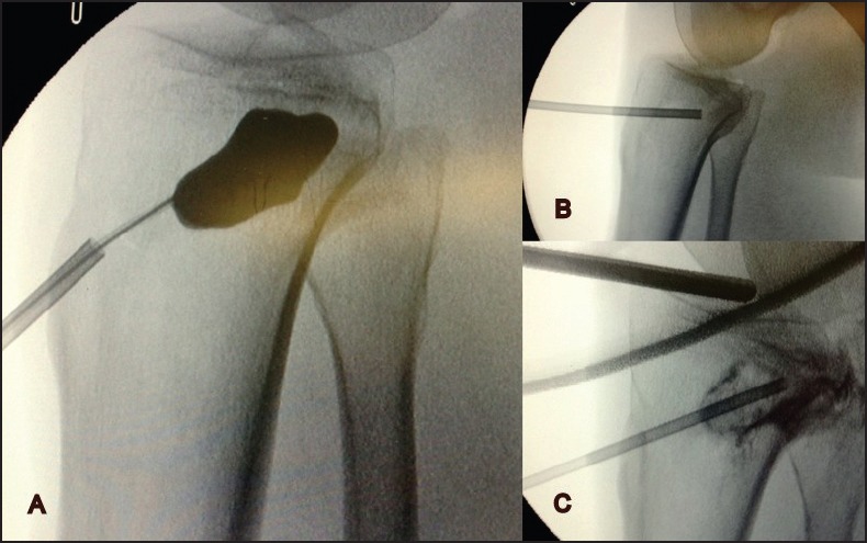
Fluoroscopy images, (a) widening of the bone defect at the fracture site using balloon, (b) proper placement of the trochar under the articular surface, (c) the cancellous cavity ready to be filled up with calcium phosphate
Official websites use .gov
A
.gov website belongs to an official
government organization in the United States.
Secure .gov websites use HTTPS
A lock (
) or https:// means you've safely
connected to the .gov website. Share sensitive
information only on official, secure websites.
Fluoroscopy images, (a) widening of the bone defect at the fracture site using balloon, (b) proper placement of the trochar under the articular surface, (c) the cancellous cavity ready to be filled up with calcium phosphate