Figure 5.

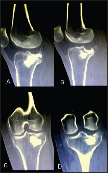
12w CT scan imaging, (a) and (b) sagittal views showing the support and maintenance of the reduction and (c), (d) coronal views showing the articular surface which is in anatomical position

12w CT scan imaging, (a) and (b) sagittal views showing the support and maintenance of the reduction and (c), (d) coronal views showing the articular surface which is in anatomical position