Figure 1.

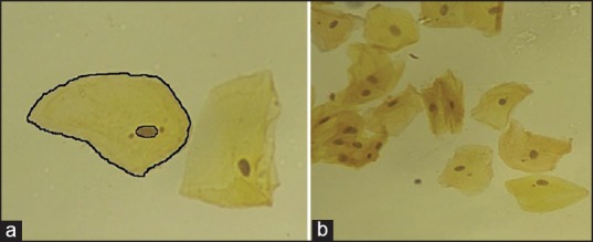
(a) Nuclear and cellular outline marked for cytomorphometry. (b) Cytological smear of normal oral mucosa stained with Papanicolaou stain (×40)

(a) Nuclear and cellular outline marked for cytomorphometry. (b) Cytological smear of normal oral mucosa stained with Papanicolaou stain (×40)