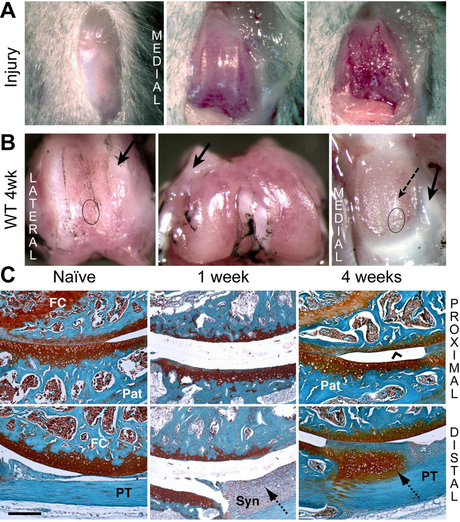
Figure 1. Macroscopic and histological evaluation of joint tissue response to cartilage injury in wild type mice.
A) The joint capsule was opened via medial peri-patellar incision, and cartilage debride d along the patellar groove of the right knee. B) The macroscopic appearance of cartilage surfaces and adjacent soft tissues was examined in the injured joint at 4 weeks post-surgery. Dense fibrous tissue formation (solid arrows) at the margins of the injured area and cartilage wear on the patella (dashed arrow) are indicated. C) Safranin-O staining of tissue structures in the femoral-patellar compartment of naïve joints and at 1 and 4 week post-injury permitted histological evaluation. The approximate regions of the groove and the patella taken for histology are indicated by circles in panel B. Cartilage resurfacing (chevron arrowhead) is observed on the femoral groove. The hyperplastic tissue deposition at the synovium (dotted arrows) at week 1 developed into a chondrophytic deposit by week 4. (Pat = patella; FC = femoral condyle; PT = patellar tendon; Syn = synovium). Scale bar = 100 μm.

