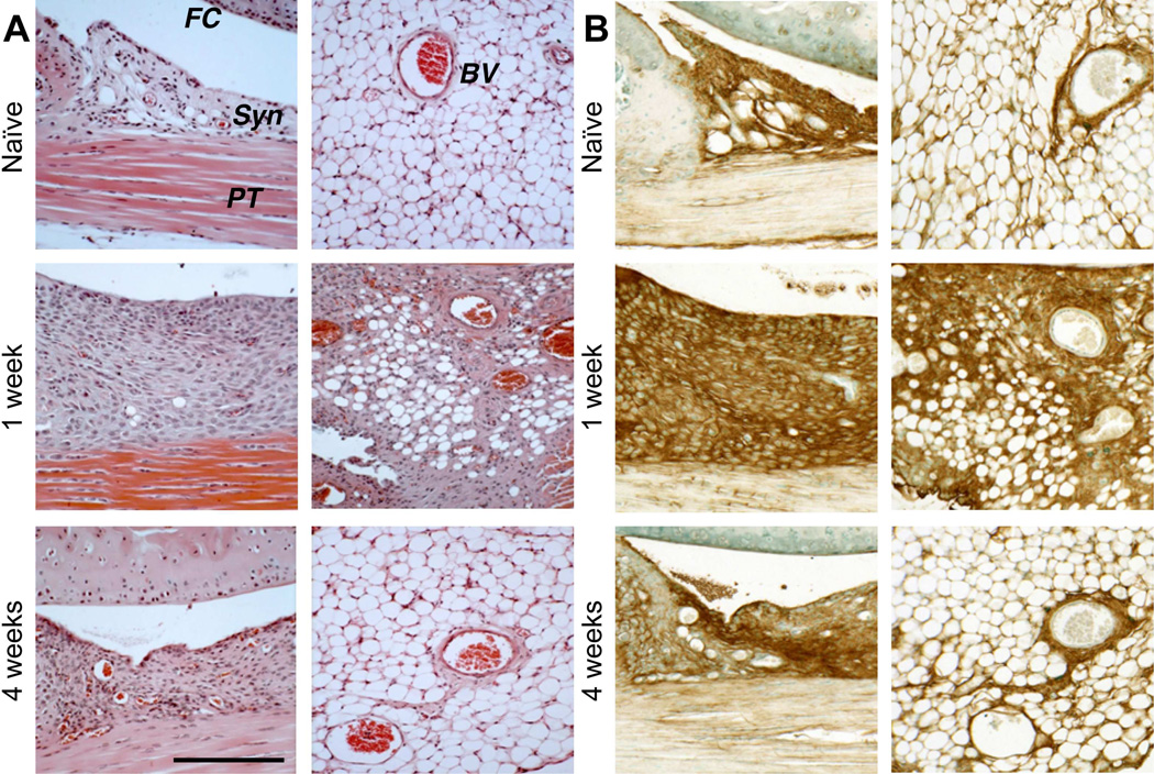
Figure 2. Histological evaluation of hyperplasia in peripatellar and perimeniscal synovium and adipose tissue following cartilage injury in wild type mice.
A) H&E staining showed post-injury cellular hyperplasia and increased ECM deposition at the synovial lining, in the adipose stroma, and perivascular regions. B) Sections equivalent to those shown in (A) were stained for HA using bHABP. (FC = femoral condyle; PT = patellar tendon; Syn = synovium; BV = blood vessel). Scale bars = 100 μm.

