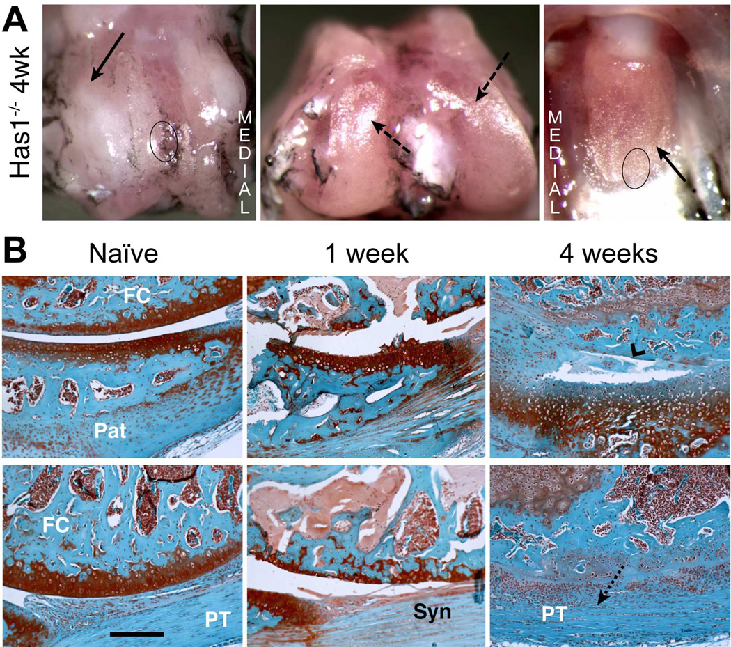
Figure 3. Macroscopic and histological evaluation of joint tissue response to cartilage injury in Has1−/− mice.
A) The macroscopic appearance of cartilage surfaces and adjacent soft tissues in the injured joint was examined at 4 weeks post-surgery. Extensive fibrotic deposits (solid arrow) were observed at the groove, and both the condylar and patellar cartilage showed signs of wear (dashed arrows). B) S ections from the femoral-patellar compartment of naïve joints and at 1 and 4 weeks post-injury were stained with Safranin-O. The approximate region of the groove and the patella taken for histology are indicated by circles in panel B. By 4 weeks post-injury, severe cartilage and bone loss (chevron arrowhead) combined with extensive fibrotic overgrowth (dotted arrow) results in a loss of joint space. (Pat = patella; FC = femoral condyle; PT = patellar tendon; Syn = synovium). Scale bar = 100 μm.

