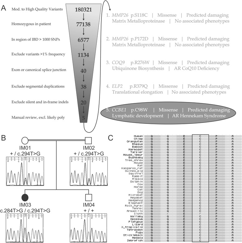
Fig. 1.

Molecular analysis identifying a mutation in CCBE1 as the likely cause of disease. (a) Exome sequencing data from patient IM03. Filter criteria are as shown. The candidates eliminated on manual review were largely known polymorphisms that were not called appropriately by the annotation algorithm. Five candidates could not be definitively excluded. The most likely candidate for the disease-causing mutation is in CCBE1 given the known function of the gene and its association with a related phenotype in humans (Hennekam Syndrome). (b) Confirmatory Sanger sequencing of the CCBE1 variant identified through the exome analysis. The variant is a T > G transversion at position 294 of the coding sequence (c.294 T > G). This changes codon 98 from a cysteine to a tryptophan (p.C98W). In the above figure, + represents wild type sequence. On the electropherograms the upper portion represents the actual sequence read and the lower portion represents the expected sequence along with the translation. (c) Multiple protein alignment with CCBE1 and its orthologs (reverse orientation). The affected amino acid is within the rectangle. This cysteine is highly conserved amongst different organisms and the mutation is predicted to be deleterious to CCBE1 function
