Fig. 3.

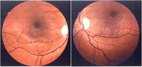
Fundus photographs of the right eye (left image) and left eye (right image). Abnormal pigmentation around the macula and mild bilateral optic atrophy can be seen.

Fundus photographs of the right eye (left image) and left eye (right image). Abnormal pigmentation around the macula and mild bilateral optic atrophy can be seen.