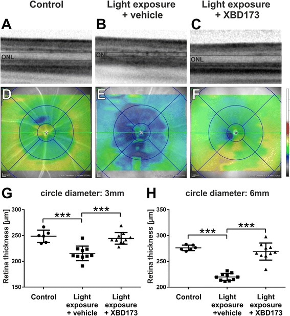
Fig. 4.

XBD173 preserves retinal thickness in light-exposed mice. SD-OCT was performed 4 days after light exposure to analyze changes in retinal structures. a–c Light-exposed mice show an altered reflectance in the ONL, which was not present in XBD173-treated mice. d–f Representative heat maps show the average retinal thickness of control and light-exposed mice after vehicle or XBD173 treatment, respectively. Light-exposed mice show a significant thinning of the retina, in the central (g) and more peripheral area (h), which was preserved by XBD173 treatment. g, h One data point represents the average thickness of the central retina, calculated from four different areas around the optic nerve head in circle diameters of 3 mm (g) and 6 mm (h), respectively. Data show mean ± SEM out of two independent experiments (control n = 6, light exposure plus vehicle treatment n = 10, light exposure plus XBD173 treatment n = 10/group) with ***p < 0.001
