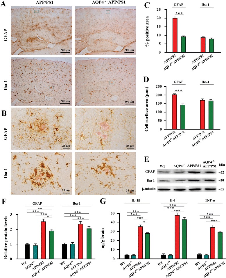
Fig. 4.

AQP4 deficiency increased astrocyte atrophy with attenuation of reactive gliosis and neuroinflammation in 12 month-old APP/PS1 mice. a GFAP positive reactive astrogliosis was significantly attenuated in the hippocampus and cerebral cortex of AQP4−/−APP/PS1 mice compared with APP/PS1 controls. A mild reduction of iba-1 positive reactive microgliosis also occurred in AQP4−/−APP/PS1 mouse brain. b The high magnification images showing that Congo Red positive plaques were surrounded by activated GFAP-positive astrocytes, displaying morphological features of hypertrophy, in APP/PS1 mice. In contrast, astrocytes adjacent to plaques exhibited atrophy with weak GFAP expression in AQP4−/−APP/PS1 mice. The reactive microgliosis around plaques was mildly reduced in AQP4−/−APP/PS1 mice, compared that in APP/PS1 controls. c The percentage of GFAP or Iba-1 positive area in the hippocampus and cortex. d GFAP-positive surface area per astrocyte and Iba-1-positive surface area per microglial cell surrounding Aβ plaques within the cortex. e Representative bands of Western bolt and f densitometry analysis of GFAP and Iba-1 protein levels in the hippocampus and cortex. g ELISA analysis of IL-1β, IL-6 and TNF-α levels in the hippocampus and cortex. Data represent mean ± SEM from 5 to 6 mice (3–4 female, and 1–2 male) per group. Data in Fig. 4c and d were analyzed by Student’s t-test and in Fig. 4f and g by ANOVA with post hoc Student-Newman-Keuls test. *P < 0.05; **P < 0.01; ***P < 0.001
