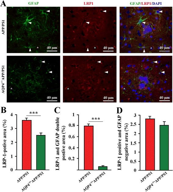
Fig. 5.

AQP4 deficiency decreased LRP1 expression by astrocytes surrounding Aβ plaques in APP/PS1 mice. a Activated GFAP-positive (green) astrocytes surrounding plaque-like structures (blue) clearly expressed LRP1 (red) (arrowheads) in 12-month old APP/PS1 mice. In contrast, in age-matching AQP4−/−APP/PS1 mice, GFAP-positive astrocytes around plaques were atrophy with very low immunofluorescent staining of LRP1 (arrowheads). Some cells negative for GFAP (stars), expressed LRP1 in the both gene-type brains. b The percentage of LRP1 positive area within a radius of 100 μm to the plaque border. c The percentage of LRP1 and GFAP double positive area. d The percentage of LRP1 positive, but GFAP negative area. Data represent mean ± SEM from 5 to 6 mice (3–4 female, and 1–2 male) per group. The statistical analysis was performed by Student’s t-test. ***P < 0.001
