Fig. 8.

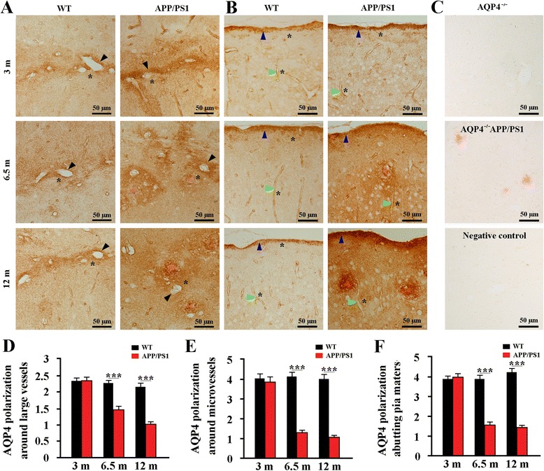
AQP4 polarization was lost during the pathological progression of APP/PS1 mice. Representative AQP4 immunoreactive images of the hippocampus a andcerebral cortex b of WT mice and APP/PS1 mice at age of 3, 6.5 and 12 months. From 3 to 12 months of age, WT mice showed high AQP4 expression closely abutting the large vessels (black arrows), microvessels (green arrows) and pia surface (blue arrows), but low AQP4 expression in the adjacent parenchyma (indicated by stars). In 3-month-old APP/PS1 mice that had no Aβ plaque disposition, AQP4 expression was slightly increased at the regions immediately abutting large vessels, microvessels and pia maters as well as correspondingly adjacent parenchymal domains. At 6.5 and 12 months of age, AQP4 was markedly increased at parenchymal domains surrounding Aβ plaques, but not significantly changed at the perivascular domains. c There was no immunoreactive signal for AQP4 in the hippocampus of AQP4−/− mice and AQP4−/−APP/PS1 mice. Brain sections from WT mice were also immuno-negative when the AQP4 antibody were omitted and replaced with an equivalent concentration of normal rabbit serum. Congo Red positive plaques were only observed in the AQP4−/−APP/PS1 hippocampus. Quantitative analyses of the AQP4 polarization abutting large vessels d (n = 16–20 per mouse), microvessels e (n = 16–20 per mouse) and pia maters f (n = 4 per mouse). Data represent mean ± SEM from 5 to 6 mice (3–4 female, and 1–2 male) per group. The statistical analysis was performed by Student’s t-test. ***P < 0.001
