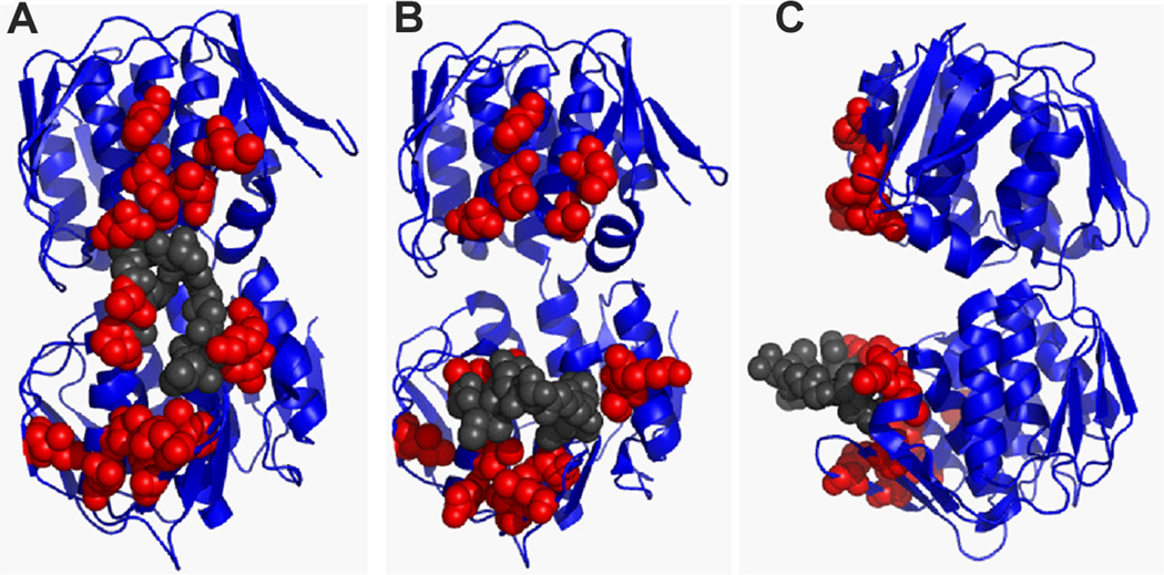
Figure 7. Rat residues define a Qβ A2 interaction surface important for inhibition of MurA.
(A) Rat residues highlighted on the MurA UDP-NAG-bound state (closed conformation, front view) (Han H., unpublished; PDB entry 3KQJ) defines a continuous surface for A2 interaction. The structure of (B) MurA unliganded (front view) (Schönbrunn et al., 2000) and (C) MurA unliganded (side view) disrupts the A2 interaction surface with the conformational change in the catalytic loop backbone. Rat residues depicted as red spheres. Residues of the catalytic loop are shown as grey spheres. Figures were generated using PyMOL (Schrödinger, 2010).

