Figure 5.
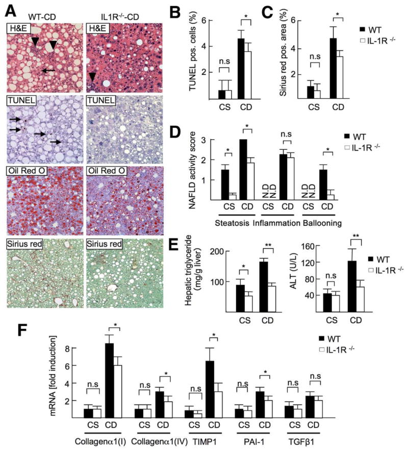
IL-1R−/− mice show attenuated steatosis and fibrosis. WT and IL-1R−/− mice were fed a CSAA diet (CS; n = 4) or CDAA diet (CD; n = 8) for 22 weeks. Closed bar, WT mice; open bar, IL-1R−/− mice. (A) H&E (arrowheads, inflammatory cells; arrows, ballooning hepatocytes), Oil-Red O, TUNEL (arrows; apoptotic cells), and Sirius red staining were performed. Original magnification, ×200 for H&E and TUNEL, ×100 for Oil-Red O and Sirius red. (B) Number of TUNEL-positive cells and (C) Sirius red–positive area were suppressed in IL-1R−/− mice. (D) NAFLD activity score, steatosis, and fibrosis were suppressed in IL-1R−/− mice. N.D., not detected. (E) Hepatic triglyceride and serum ALT levels were decreased in IL-1R−/− mice. (F) Hepatic mRNA levels of fibrogenic markers were measured by quantitative real-time PCR. Data represent mean ± SD, *P < .05; n.s., not significant.

