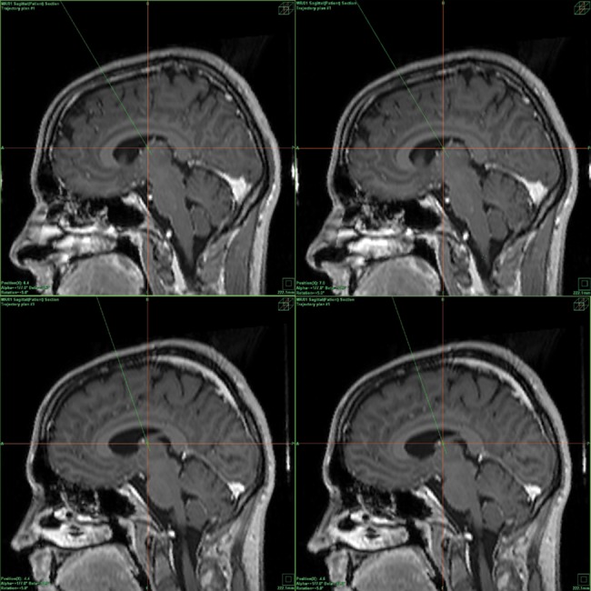
Fig 1. Example of the actual positions of the all ANT contacts in patient 2 (right-left orientation of the sagittal scans, fusion of the MR scans and CT correlation).

Upper picture: position of the right ANT contacts: the R3 contact (left picture), and position of the R4 contact (right picture). Lower picture: position of the left ANT contacts: the L3 contact (left picture) and position of the L4 contact (right picture).
