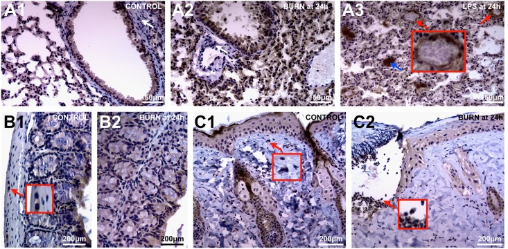
Fig 3. CHI3L1-expression in tissues is dominated by the epithelial lineage as well as a small subset of stromal cells.
Immunohistochemistry using anti-CHI3L1 was performed in WT mice. (A1) Both bronchial and alveolar epithelium stain CHI3L1+ in normal lung tissue. (A2) With burn-induced lung injury peri-vascular and–bronchial stromal tissue remains CHI3L1- (white arrow). However, with (A3) severe, LPS-induced SIR, lung tissue demonstrates CHI3L1+ vascular endothelium (red arrow) as well as CHI3L1+ mucous bronchial deposits (blue arrow). (B1) IHC confirms constitutive CHI3L1+ epithelium in colon along with few stromal cells (red arrow) apparently not affected by (B2) burn injury. (C1) Along with epithelium, CHI3L1+ stromal cells are also present in skin (red arrow). (C2) With burn injury, CHI3L1+ granulocytes accumulate at the zone of demarcation.

