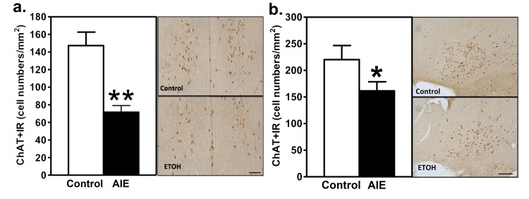
Fig 3. AIE decreases ChAT+IR in the basal forebrain in adulthood.
AIE decreases ChAT+IR in the Ch1 and Ch2 nuclei of the basal forebrain of adult rats (a). Left Panel—the cell density of ChAT+IR is significantly decreased in the Ch1 and Ch2 nuclei at 0.70 ~ 0.20 mm from bregma 25 days after AIE, **p<0.01. Right Panel—representative photomicrography ChAT+IR neurons in the Ch1 and Ch2 nuclei from a control animal (Control), and an AIE-exposed animal (ETOH). AIE decreases ChAT+IR in the Ch3 and Ch4 nuclei of the basal forebrain of adult rats (b). Left Panel—the cell density of ChAT+IR is significantly decreased in the Ch3 and Ch4 nuclei at 0.48 ~ 0.40 mm from bregma 25 days after AIE, *p = 0.046. Right Panel—representative photomicrography ChAT+IR neurons in the Ch3 and Ch4 nuclei from a control animal (Control), and an ethanol-exposed animal (ETOH). Scale bar = 50 μm.

