
Extended Data Figure 8. Karyotyping analysis and pluripotency of newly generated iPSC lines.
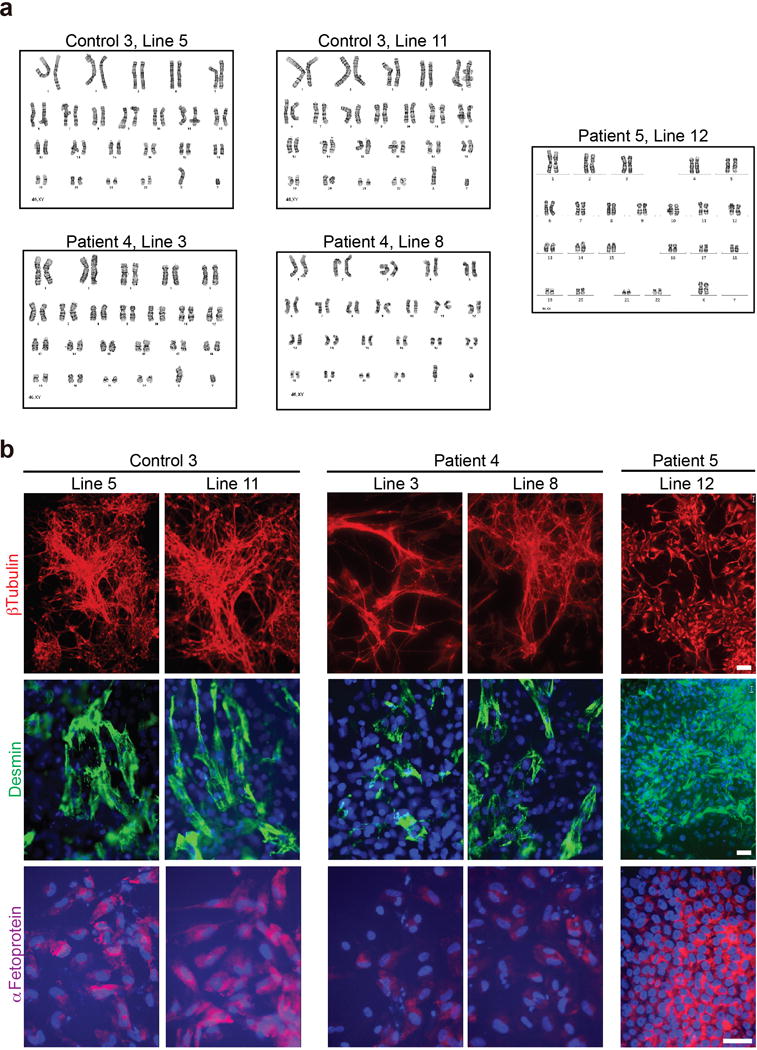
a, G-band staining showing a normal karyotype for all the lines analyzed. b, After in vitro spontaneous differentiation of control and C9 carrier iPSC lines, cells were staining for α-fetoprotein (AFP, endoderm), desmin (mesoderm), βIII-tubulin (ectoderm), and Hoechst (nuclei). All lines showed differentiation towards derivates of three germ layers. Scale bars: 20 mm.