
Fig. 4.
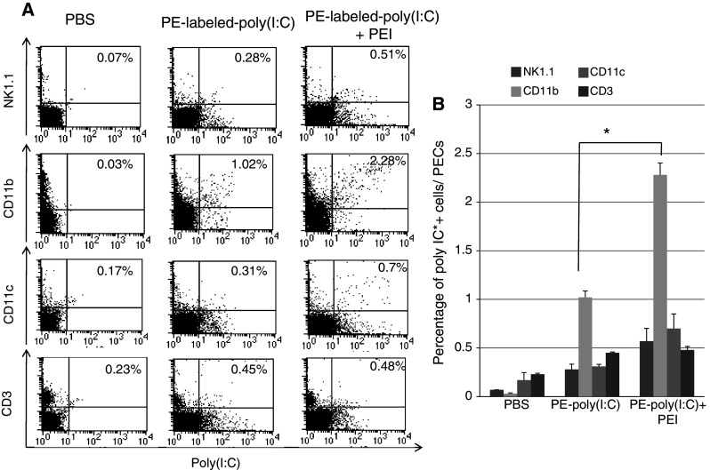
Flow cytometry analysis to determine the uptake of PE-labeled poly(I:C) into various types of peritoneal cells with or without PEI. Groups of C57BL/6 mice were i.p. injected with PBS, PE-labeled-poly(I:C) (1 μg/mouse) alone or in combination with PEI (0.16 μl/mouse, N/P = 8). PECs were harvested 16 h after treatment and stained for NK1.1, CD11b, CD11c and CD3 surface markers and analyzed by flow cytometry analysis. a Representative flow cytometry data demonstrating the percentage of NK1.1, CD11b, CD11c and CD3+ cells transfected with poly(I:C) among all PECs (right upper quadrant). Significant amounts of CD11b+ cells (5.07% of CD11b+ cells and 2.28% of all PECs) were positive for poly(I:C). b Bar graph depicting the percentage of poly(I:C)+ cells among all PECs. Data are represented as mean ± SD. *indicates P < 0.05. Data shown are representative of two experiments conducted