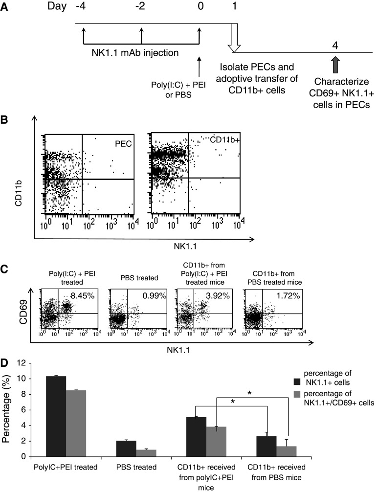
Fig. 6.
Characterization of the number of CD69 + NK1.1+ cells in the PECs of mice receiving adoptive transfer of CD11b+ cells from mice treated with or without poly(I:C) and PEI. a Schematic diagram to depict the depletion of NK1.1, treatment regimen and adoptive transfer of CD11b+ cells in mice. Naïve C57BL/6 mice were treated with PBS or combined treatment (poly(I:C)+ PEI) on day 0. Mice were depleted of NK1.1 cells using PK136 Ab injected i.p. 3 times every other day, 4 days before treatment. On day 1 after treatment, PECs were harvested and CD11b+ cells were isolated; 5 × 10⁵ of isolated CD11b+ cells were then adoptively transferred i.p. into a new group of naïve C57BL/6 mice. Three days later, PECs from the recipient mice were collected and analyzed for the number of total NK1.1+ cells and CD69 + NK1.1+ cells using flow cytometry. b Representative flow cytometry data to characterize the isolated NK1.1-CD11b+ in the PECs. c Representative flow cytometry data demonstrating the percentage of CD69 + NK1.1+ cells following adoptive transfer of CD11b+ cells from mice treated with PBS (upper panel) or poly(I:C) + PEI (lower panel). d Bar graph depicting the percentage of total NK1.1+ cells and CD69 + NK1.1+ cells among all PECs following adoptive transfer of CD11b+ cells from mice treated with poly(I:C) + PEI or PBS. *indicates P < 0.05. Data shown are representative of two experiments conducted

