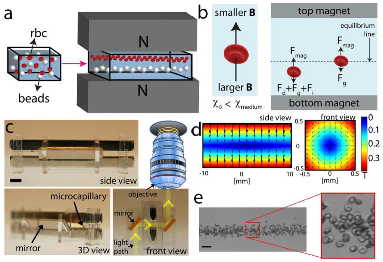
Figure 1. Magnetic levitation-based device for static separation, cytometry and functional interrogation of cells.
(a) Schematic of the magnetic levitation and cell manipulation in a magnetic field. (b) Negative difference between the magnetic susceptibilities of an object (χ0) and suspending medium (χmedium) causes the object to move away from larger magnetic field strength site to lower magnetic field strength. Until the object reaches the equilibrium, fluidic drag (Fd), inertial (Fi), buoyancy (Fg), and magnetic forces (Fm) act on the object. As the object gets closer to the equilibrium, its migration velocity, and thus drug and inertial forces, become smaller. (c) Front, 3D, and side views of the magnetic levitation setup. A capillary with 1 mm × 1 mm square cross-section is placed between two permanent neodymium magnets (NdFeB) in a configuration where same poles facing each other. Scale bar is 3 mm. (d) Controlling the position and orientation of objects with magnetic field gradients. Contour plot of the magnetic field for two magnets with same poles facing each other. The magnitude of the magnetic field is constrained between 0 T and 0.4 T. More information on modeling is given in Supplementary Information. (e) Confinement of RBCs in 50 mM Gd+ solution. Scale bar is 40 μm.

