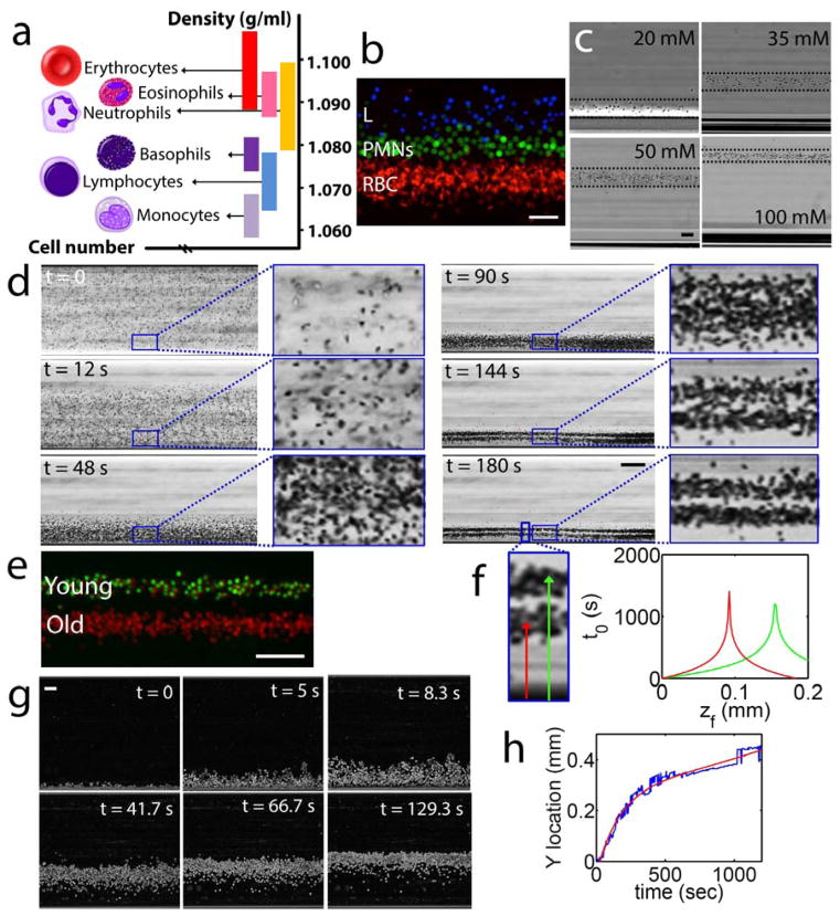
Figure 2. Characterization of cell separation using magnetic levitation.
(a) Density histogram of monocytes, lymphocytes, basophils, PMNs, eosinophils, and RBCs. (b) Magnetically-driven, density-based separation of fluorescently-labeled blood cells. Freshly purified RBCs (RBCs, labeled red), PMNs (PMNs, labeled green), and lymphocytes (L, labeled blue) were mixed and magnetically confined for 15 minutes in 30 mM Gd+ solution. Scale bar is 40 μm. (c) Levitation of RBCs at different molarities of suspending Gd+ solution, from left to right: 20, 35, 50, and 100 mM Gd+ solution. Scale bar is 40 μm. (d) Separation of young and old RBCs by age (Supplementary Movie 1). Time-lapse images showing the density separation of a mixed population of purified young and old RBCs. Scale bar is 160 μm. (e) Fluorescently labeled young (green) and old (red) RBCs at their equilibrium levitation height. Scale bar is 100 μm. (f) Analytical equilibration time as a function of equilibration height of old and young RBCs. (g) True magnetic levitation in whole blood (diluted 1:1000). Sedimented RBCs were loaded into the magnetic levitation device and cells imaged for 20 minutes. Scale bar is 40 μm. (h) Time-dependent location of RBCs levitating from bottom surface of microcapillary toward their equilibration point.

