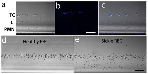
Figure 4. Versatility of magnetic levitation-based approach.

(a–c) Identification of circulating tumor cells in whole blood. Diluted blood isolated from a healthy donor was spiked with breast cancer cells (TC) and magnetically confined using a low (15 mM) Gd+ concentration that allows only levitation of TC, PMNs (PMN) and lymphocytes (L) but not that of RBCs. (a) Brightfield image of the levitated cells. (b) Fluorescent image of the levitated cells. TCs are labelled with blue. (e) Merged image of brightfield and fluorescent image. Scale bar is 100 μm. (d&e) Identification of sickle cell disease by magnetic levitation. Levitation of (d) healthy and (e) sickle RBCs in the presence of 10 mM Na metabisulfite. Bright filed images of RBCs recorded after 10 minutes of confinement. Enhanced (Roberts filter), image analysis-friendly images of the same snapshots are given in Fig. S6. Scale bar is 100 μm.
