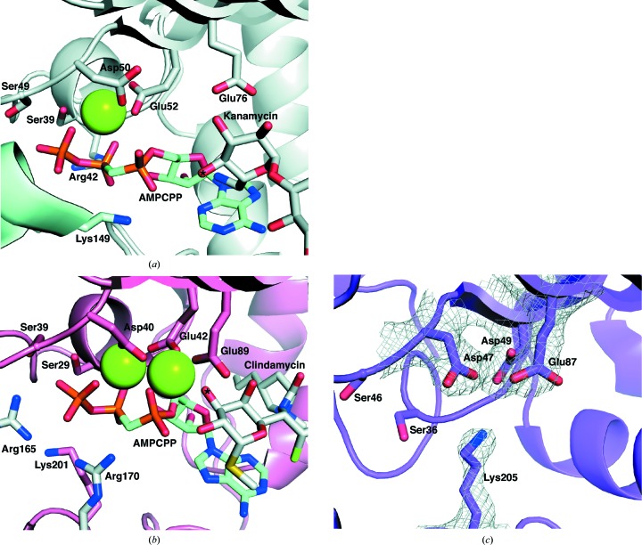
Figure 5.
Nucleotide-binding sites of KNTase (a), LinB (b) and AadA (c). All figures are in the same orientation, according to superpositions based on the N-terminal domain. Mg2+ ions are shown as green spheres. A 2F o − F c map for Asp47, Asp49, Glu87 and Lys205 in AadA is shown in (c) contoured at 1σ (0.23 e Å−3). The equivalent residues in KNTase and LinB are shown in (a) and (b). The adenylation sites of kanamycin and clindamycin are indicated by asterisks. The two monomers of KNTase (a) are shown in grey and pale blue and the two monomers of LinB (b) are shown in salmon and grey.

