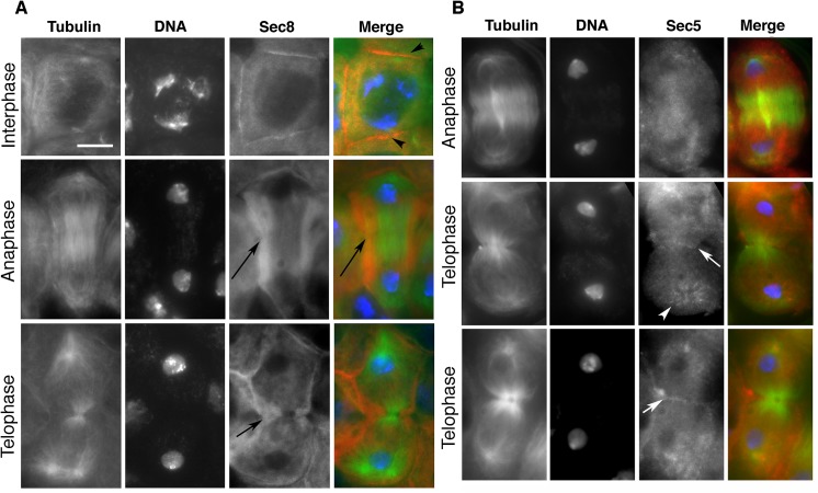
Fig 2. Localization of exocyst complex proteins in dividing spermatocytes.
(A) Localization of Sec8 protein in wild-type primary spermatocytes. Interphase and dividing spermatocytes were stained for Tubulin (green), Sec8 (red) and DNA (blue). During interphase, Sec8 was mostly diffuse in the cytoplasm and enriched at the plasma membrane (arrowheads). In dividing spermatocytes, Sec8 appeared enriched in a broad cortical band that encircled the midzone (arrows) and was excluded from the poles. (B) Localization of Sec5 protein in wild-type dividing spermatocytes. Primary spermatocytes were stained for Tubulin (green), Sec5 (red) and DNA (blue). Note the enrichment of Sec5 in puncta at the astral microtubules (arrowhead) and at the cleavage furrow (arrows). Scale bar, 10 μm.

