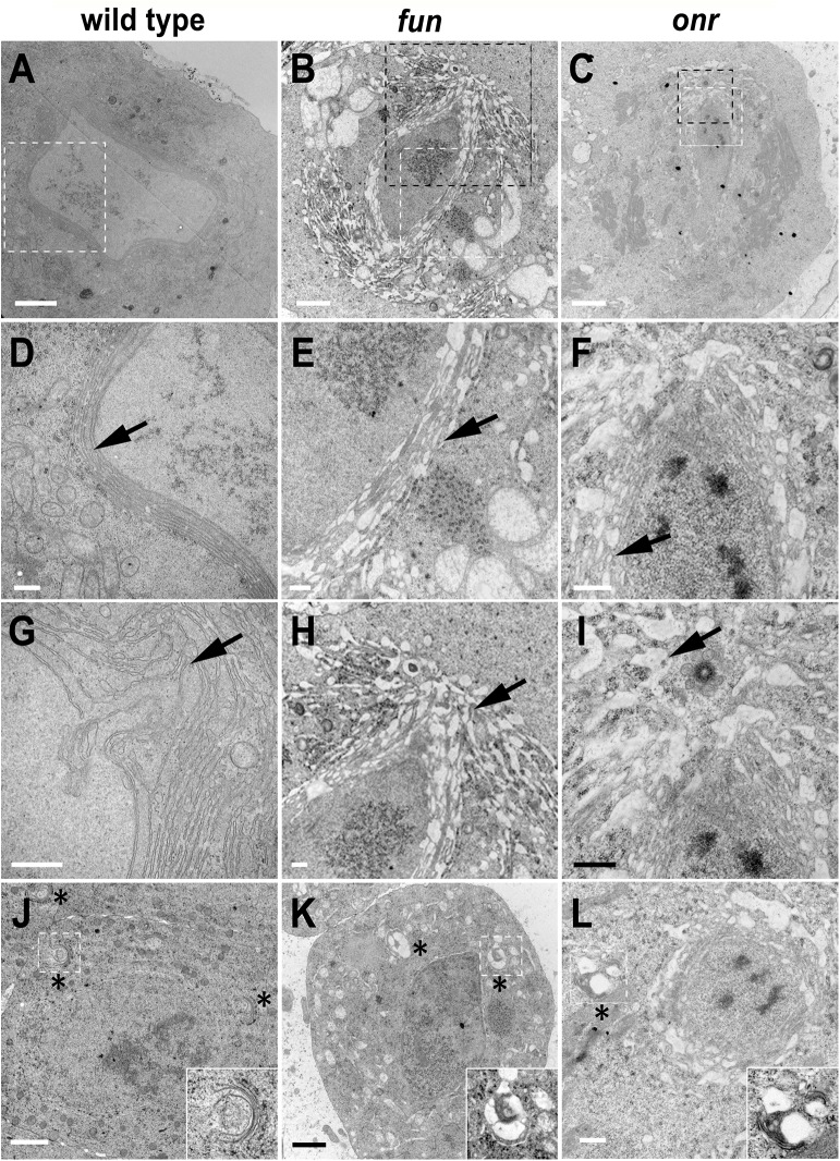
Fig 6. Defects in morphology and ultrastructure of parafusorial membranes and Golgi bodies in fun and onr mutant cells.
Transmission electron micrographs showing parafusorial membranes (A-F), astral membranes (G-I), and Golgi bodies (J-L) in fun and onr mutant spermatocytes. Parafusorial and astral membranes (arrows) are enlarged, fragmented and vacuolated in fun z1010/Df(3R)Exel6145 (B, E, H) and onr z4840 /Df(3R)Espl3 (C, F, I) dividing spermatocytes. (D, E, F) panels are magnified images of areas surrounded by white squares in (A, B, C). (H, I) panels are magnified images of areas surrounded by black squares in (B, C). Golgi bodies (asterisks) show vacuolated regions in fun (K) and onr (L) mutant spermatocytes. Golgi bodies surrounded by white squares in (J-L) are magnified in insets. Scale bars are 2 μm (A-C, J, K) or 500 nm (D-I, L).

