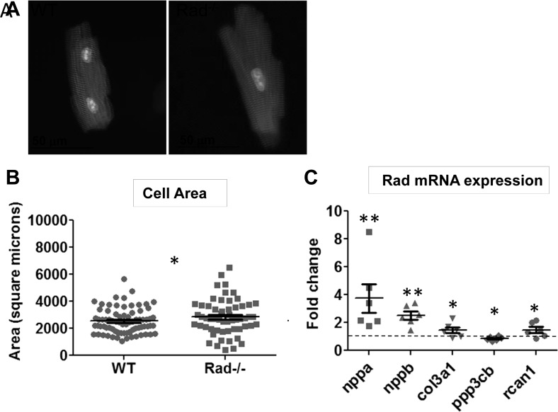
Fig. 2.
Rad deletion increases cell size and upregulates fetal and growth markers. A: representative isolated ventricular myocytes stained for α-actinin (red) and nucleus (blue). B: an increase in area is observed in myocytes isolated from Rad−/− hearts; n = 3 hearts. *P < 0.05 vs. WT. C: upregulation of hypertrophic markers nppa (ANF), nppb (BNP), collagen isoform col3a1, pp3cb (calcineurin), and rcan1 (MCIP) in Rad−/− hearts; n = 6 hearts. *P < 0.05, **P < 0.01 vs. WT.

