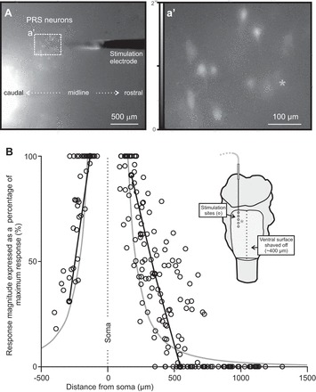
Fig. 2.

Local activation of pRS neurons by PRF stimulation. A: low-magnification image (ventral view) of a stimulation microelectrode (tip) inserted in the PRF of a brain stem preparation for which the ventral surface had been shaved and in which pRS neurons (and other spinally projecting neurons) were retrogradely labeled with CGDA. The microelectrode entered the tissue ∼750 μm rostral to the rostral edge of the field of view, which shows the CGDA-labeled pRS neurons from which calcium recordings were obtained (dotted rectangle). a′: High-magnification image (single frame) showing baseline calcium fluorescence in some of the CGDA-labeled pRS neurons surrounding the microelectrode tip shown in A. A gray asterisk is placed above the soma of pRS neuron 3 in this experiment. The same neuron is singled out in B as well (black lines in graph and gray asterisk in schematic). B: relationship between response magnitude and distance from microelectrode tip for 38 pRS neurons (4 animals, 1–2 electrode tracks per animal). For each pRS neuron, the magnitudes of responses to a 2-s stimulus train of 120 μA at 10 Hz (1 response for each site stimulated along the electrode track) are plotted as a percentage of the largest response evoked in that neuron. The distances between pRS somata and stimulation sites in 3-dimensional space are calculated with basic trigonometry using distances obtained from overview pictures similar to the 1 shown in A and histological assessments of the stimulation sites (see methods). The 2 black lines join the responses of pRS neuron 3 evoked at stimulation sites 1–3 and 4–5, respectively. The similarly orderly behavior of the other pRS neurons is concealed in the broad point-cloud distribution. Solid gray curves follow a function that describes the inverse of the squared distance (see text). The inset shows a cartoon of the experimental setup with the approximate position of pRS neuron 3 (gray asterisk) relative to the 5 sites stimulated along the electrode track (open circles).
