
Fig. 6.
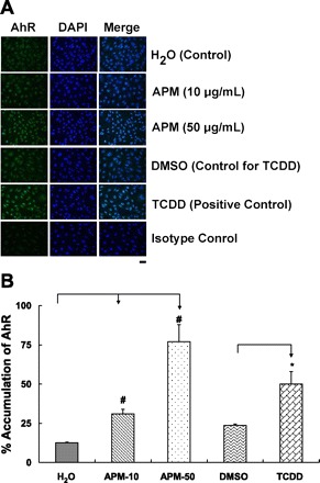
A: aryl hydrocarbon receptor (AhR) nuclear localization in HAEC treated with Sum06 APM for 1 h. Representative images of HAEC monolayers showing AhR distribution in cytosol and nucleus. Nuclear accumulation of AhR was evident after exposure to sum06 APM or tetrachlorodibenzo-p-dioxin (TCDD, positive control) compared with control cells treated with H2O or DMSO (control for TCDD) by immunofluorescence staining (n = 4). Isotype controls had no detectable staining. Bar = 40 μm. B: percent accumulation of AhR by sum06 APM. Percent positive nuclei after 1 h incubation. 31% (APM-10), 77% (APM-50), and 50% (1 nM TCDD) of nuclei stained with AhR after 1 h. APM exposure compared with control H2O and TCDD was compared with control DMSO. n = 4; *P ≤ 0.05; #P ≤ 0.005.