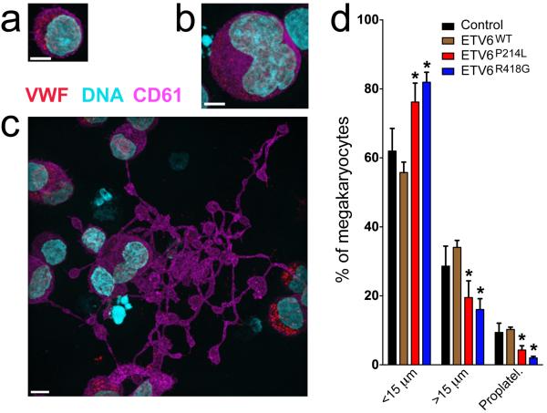
Figure 2.
Abnormal development of day 12 cultured megakaryocytes expressing mutant ETV6. Control cells and those transduced with lentivirus to express myc-tagged forms of ETV6 (ETV6WT, ETV6P214L and ETV6R418G) were assessed via immunofluorescence microscopy imaging; scale bars = 5 μm. (a–c) Megakaryocytes (control cells shown) were identified via expression of CD61 (violet) and VWF (red) and staged by diameter: <15 μm (a), >15μm (b), or the presence of proplatelets (c). (d) Population distributions showed no significant differences between control and ETV6WT-transduced cells, while ETV6P214L-transduced megakaryocytes showed a significantly higher proportion (*P<0.05, 2-tailed t-test) of cells in the earlier developmental stage (<15 μm) and fewer in the mature proplatelet-forming stage (control: n=7, WT: n=3, P214L: n=3, R418G: n=4 cultures with >300 cells for each; error bars = standard deviation). Images for control cells and those transduced with WT, P214L and R418G forms of ETV6 are shown in Supplementary Fig 6.

